Cross Section Olfactory Organ Nasal Cavity Vomeronasal Organ Marsh Frog — 스톡 사진

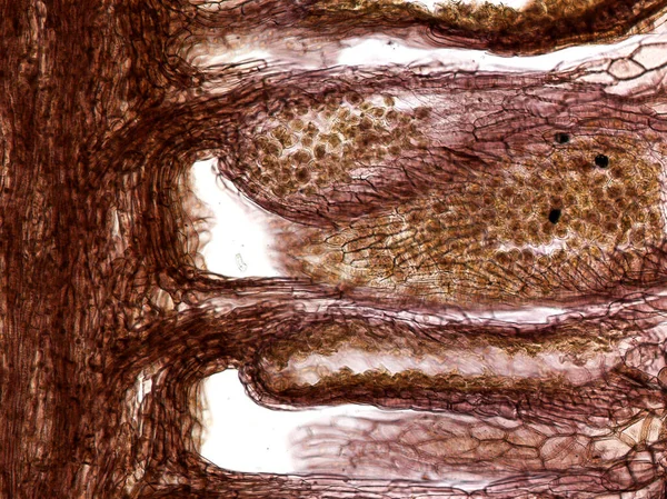
Cross section of the olfactory organ (nasal cavity, vomeronasal organ) of the marsh frog (Pelophylax ridibundus). Hematoxylin and Eosin Staining (H&E stain). — 스톡 이미지

Cross section of the olfactory organ (nasal cavity, vomeronasal organ) of the marsh frog (Pelophylax ridibundus). Hematoxylin and Eosin Staining (H&E stain).

 — 님의 사진 BioFoto

동일 시리즈:

Trachea, light micrograph. 기관 (氣管) 으로부터 의 아드레날린 연골 , 콜라겐 섬유 및 매트릭스 를 보여 주는 것.
Trachea Light Micrograph 으로부터 아드레날린 콜라겐 매트릭스 — 스톡 사진
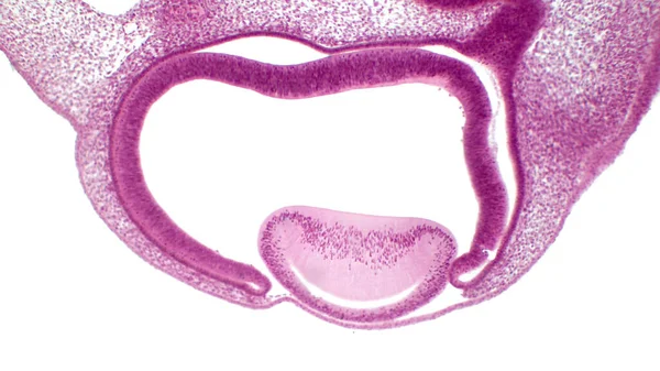
Eye development. Cells from both the mesodermal and the ectodermal tissues contribute to the formation of the eye. In the center is a crystalline lens.
Eye Development Cells Both Mesodermal Ectodermal Tissues Contribute Formation Eye — 스톡 사진
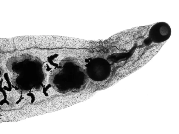
소 (小) 소 (小) 포구 (小球) 와 꽃가루를 보여 주는 남성 소나무 (Pinus sylvestris) 의가늘고 긴 부분.
꽃가루를 소나무 Pinus Sylvestris 의가늘고 — 스톡 사진
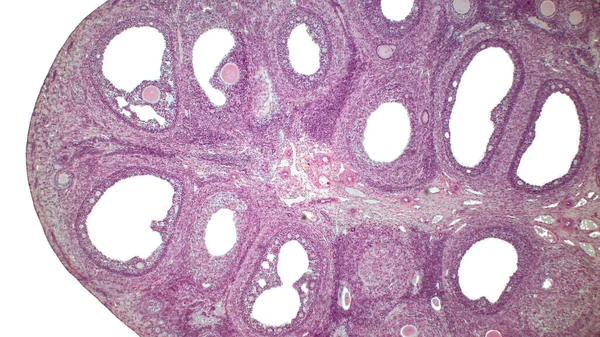
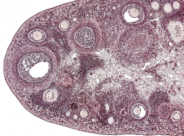
Light micrograph of the Cat's ovarian. Micrograph of ovary showing primordial, primary and secondary follicles isolated on white background. Hematoxylin and eosin stain.
Light Micrograph Cat Ovarian Micrograph Ovary Showing Primordial Primary Secondary — 스톡 사진
사이 불극성 신경 세포입니다. Pseudounipolar neurons of rounded soma of a dorsal root ganglion.ell body is surrounded by glia cells called satellite cells. 헤마 토크 린 과에 오 신 얼룩이야. 확대 사진 : x1000.
불극성 세포입니다 Pseudounipolar Neurons Rounded Soma Dorsal Root Ganglion Ell — 스톡 사진
소뇌의 횡단면. 광선 현미경.
소뇌의 횡단면 현미경 — 스톡 사진
입에서 기관지와 폐로 연결되는 기관 (氣管) 을 통해 기관 (氣管) 을 통과하는 부분을 미세하게 찍은 것이다. 기도 (氣道) 공동 은호 흡기 상피 골로 둘러싸여 있다.
입에서 기관지와 연결되는 통과하는 부분을 미세하게 것이다 둘러싸여 — 스톡 사진
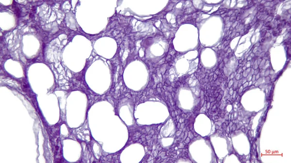
빛 현미경: 흉골의 흰 지방 조직. 커다란 밝은 구멍들 - 쇄빙선 의유해.
현미경 흉골의 커다란 구멍들 쇄빙선 의유해 — 스톡 사진
횡단면의 골격근 섬유는 세포 주변에 위치 한 여러 개의 핵 다세포 세포의 존재를 보여준다. 빛 현미경 현미경 현미경 편광계.
횡단면의 골격근 섬유는 주변에 다세포 세포의 존재를 보여준다 현미경 현미경 — 스톡 사진
소뇌의 횡단면. 광선 현미경.
소뇌의 횡단면 현미경 — 스톡 사진
소뇌의 횡단면. 광선 현미경.
소뇌의 횡단면 현미경 — 스톡 사진
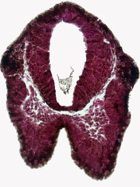
Cross section of the olfactory placode and brain of the embryo frog. Hematoxylin and Eosin Staining (H&E stain).
Cross Section Olfactory Placode Brain Embryo Frog Hematoxylin Eosin Staining — 스톡 사진
소뇌의 횡단면. 광선 현미경.
소뇌의 횡단면 현미경 — 스톡 사진
소뇌의 횡단면. 광선 현미경.
소뇌의 횡단면 현미경 — 스톡 사진
간단 한 칼럼에 피텔리움. 신장 속질 (renal medulla) 의밝은 현미경 사진은 단순 한 칼럼 상피 (epithelium) 가 줄지어 있는 여러 개의 수집 관을 보여준다.
칼럼에 피텔리움 Renal Medulla 의밝은 현미경 사진은 Epithelium 줄지어 보여준다 — 스톡 사진
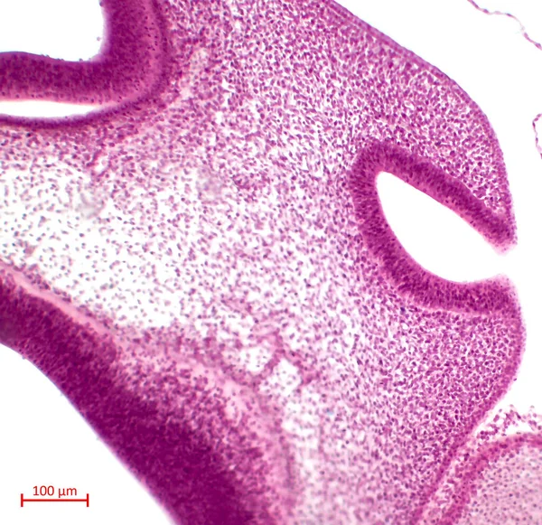
Olfactory organ development. Olfactory fossa pit stage. Hematoxylin and Eosin Staining (H&E stain).
Olfactory Organ Development Olfactory Fossa Pit Stage Hematoxylin Eosin Staining — 스톡 사진

사용 정보

표준 또는 확장 라이센스에 따라 이 로열티-프리 "Cross section of the olfactory organ (nasal cavity, vomeronasal organ) of the marsh frog (Pelophylax ridibundus). Hematoxylin and Eosin Staining (H&E stain)." 사진을 개인적 및 상업적 목적으로 사용할 수 있습니다. 표준 라이센스는 광고, UI 디자인, 제품 패키지를 비롯한 대부분의 사용을 커버하며, 최대 500,000부까지 인쇄할 수 있습니다. 확장 라이센스는 무제한 인쇄 권한과 함께 표준 라이센스 하의 모든 사용을 커버하며 다운로드한 스톡 이미지를 상품, 제품 재판매 또는 무료 배포에 사용할 수 있도록 합니다.

이 스톡 사진 구입 시 최대 2678x1766 의 고해상도로 다운로드할 수 있습니다. 업데이트 날짜: 2022년 5월 15일